
Technology — The promise of CTCs
The promise of CTCs
Circulating Tumor Cells (CTCs) are malignant cells that are shed from the primary or metastatic solid tumors and then infiltrate into the vascular and lymphatic systems; these cells play a fundamental role in the metastatic process of non-hematological cancers. Once inside the bloodstream, they can easily colonize new tissues. Moreover, they can remain dormant for many years before they form a tumor.
When cells are dormant, they can continue to evade the immune system and even chemotherapy or other forms of treatment. Because they are still subjected to the effects of the drugs, however, they can develop new mutations that make them resistant and allow them to keep proliferating.
How do they reach the bloodstream?
CTCs can enter the bloodstream actively or passively. Due to the rapid growth of tissue in the tumor, blood vessels close to the tumor become fragile and permeable. Under these circumstances, either single cells or cell clusters can cross the endothelium passively and enter the bloodstream, a process known as intravasation.
Another theory states that endothelial cells can undergo a process known as endothelial to mesenchymal transition (EMT), where they lose their adhesion capacity and, in exchange, begin expressing properties that allow them to enter the vasculature.
Some of the changes related to EMT include loss of cell polarity, disruption of cell-cell junctions and the reorganization of the ECM, as well as the down-regulation of E-cadherin, EpCAM and certain cytokeratins. On the other hand, the upregulation of N-cadherin, vimentin, fibronectin and integrin β1 and β3 indicates the induction of a mesenchymal state.
Importantly, these cells very rarely advance to a fully mesenchymal state, but rather express characteristics of both types of cells, meaning that they can reverse this transition. The re-expression of proteins specific to epithelial cells, a process known as mesenchymal to epithelial transition (MET), may be the final step by which CTCs colonize a different organ.
Why are they hard to trap?
The isolation of these malignant cells from blood represents a major technological challenge due to their heterogeneity and extremely low numbers in comparison to blood cells. Current separation methods take advantage of antigen-antibody interactions to capture CTCs for further analysis. CellSearch, the only FDA-approved device, as well as many other popular kits are based on this concept. Nevertheless, CTCs are highly heterogeneous and these conventional methods are unable to effectively isolate cells that do not express the expected biomarkers.
In addition, CTCs are extremely scarce in blood. On average you can find around 40.5 billion cells in 7.5 mL of blood, while a cancer patient may have between 1 and 1000 CTCs in the same volume. Although some methods use a preconcentration step to reduce the amount of blood cells, CTCs can be lost during that process, leading to incomplete or erroneous results.
Current isolation methods are either incompatible with whole blood samples or lack the sensitivity and specificity needed to isolate CTCs reliably, which has prevented the development of assays with real clinical utility… until now.
Why are they important?
The importance of CTCs is related to their clinical implementation. At the beginning, counting the number of cells was the main purpose of their isolation, but as knowledge regarding the metastatic cascade increased, the interest shifted towards evaluating their genomic profile. Today, we know that the number of CTCs does not relate directly to the stage of the disease, but that the receptor variations are linked to the aggressiveness of the disease.
Currently, disease status is assessed via a standard biopsy, where a small part of the tumor is directly obtained during a surgical procedure. Although the genetic information obtained from these cells is critical for treatment selection, the process is uncomfortable and risky for the patient, and in some cases impossible because the tumor is inaccessible.
Using CTCs as biomarkers overcomes many of the drawbacks of conventional tissue biopsy. It is less invasive and faster, and it better reflects the global heterogeneity of the tumor, including the metastatic sites. Furthermore, the procedure can be repeated over time to test for treatment resistance and adjust the treatment plan as soon as possible.
At the moment, CTCs are not meant to replace tissue biopsy, but rather to complement the information given by this analysis and increase the chances of responsiveness to treatment.
The CytoCatch Technology
CTCs are found in blood together with other types of cells, including erythrocytes, platelets and immune cells. One of the main characteristics of CTCs is their size. These cells are much larger and less deformable than most of the other blood components, so they can be easily separated by size.
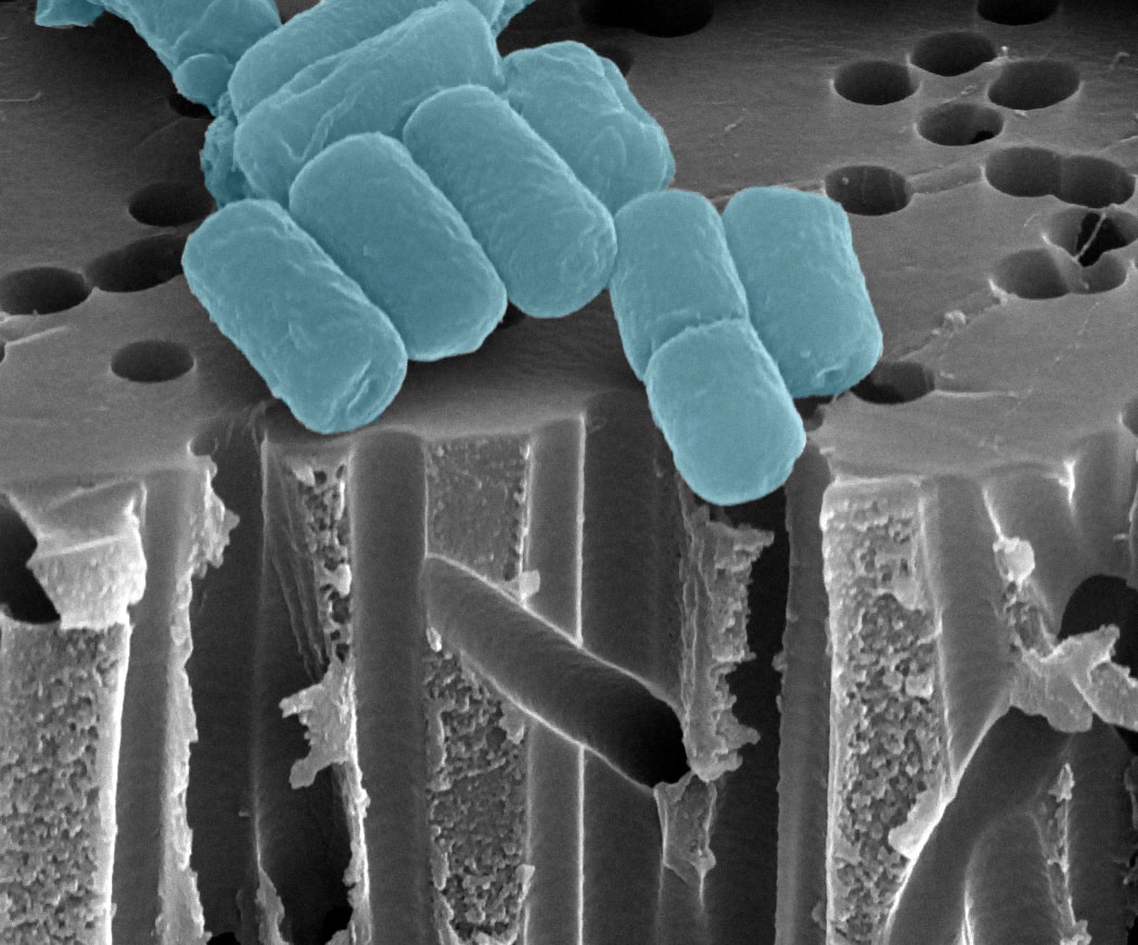
The Cytocatch™ technology is based on a filter. As the sample is pressurized and moved through the filtration membrane, the cancer cells are separated from the blood so they can be further analysed. Just as simple: no labels or extra steps required. This principle has consistently proven its effectiveness at isolating a greater number of CTCs in samples from patients with different types of cancer, even capturing CTC subtypes that no longer express EpCAM antigens, when compared with approaches based on capture antibodies.
How are we different from other techniques?
Most of the technologies developed to isolate CTCs from blood are based on sample enrichment methods that depend on specific antigen-antibody interactions. This includes the CellSearch system, considered the current gold standard. Although these technologies have demonstrated clinical utility, a fundamental problem of this approach is the lack of a universal surface marker that is consistently expressed by CTCs. This means that many cells are lost during the process, missing valuable information about the heterogeneity of the disease.
Size-based isolation overcomes this problem, showing higher capture efficiencies. However, various microfiltration technologies use polycarbonate track-etch membranes. The processes used to fabricate these membranes create random pores in the plastic sheet, making them inadequate for this end. The membranes are restricted to a low number of pores, which can lead to cell damage (due to uneven distribution of pressure over the membrane), lower capture efficiency, reduced reproducibility, and a higher recovery of non-specific cells.
Our system uses an electroformed nickel membrane. This microfabrication method not only produces a uniform hole pattern, but it is also a highly scalable process which allows us to fulfill the necessities of the medical field.
Furthermore, the shear stress exerted on the cells is minimal, so they remain viable for downstream analysis using traditional molecular biology techniques and next-generation sequencing technologies.
The CytoCatch™ Isolation Platform is capable of capturing CTC subtypes that no longer express epithelial antigens.

Assay workflow
Develop your own panel
To analyse the captured cells, the CytoCatch™ Imaging System possesses special routines and machine learning algorithms that analyze the captured cells based on their morphology and the expression of specific markers.
See the imaging systemHave questions about the technology?
Our team is happy to walk you through the science, the hardware, and what it takes to run the CytoCatch™ platform in your lab.