
Cytocatch Imaging System


Enabling high performance isolation of CTCs
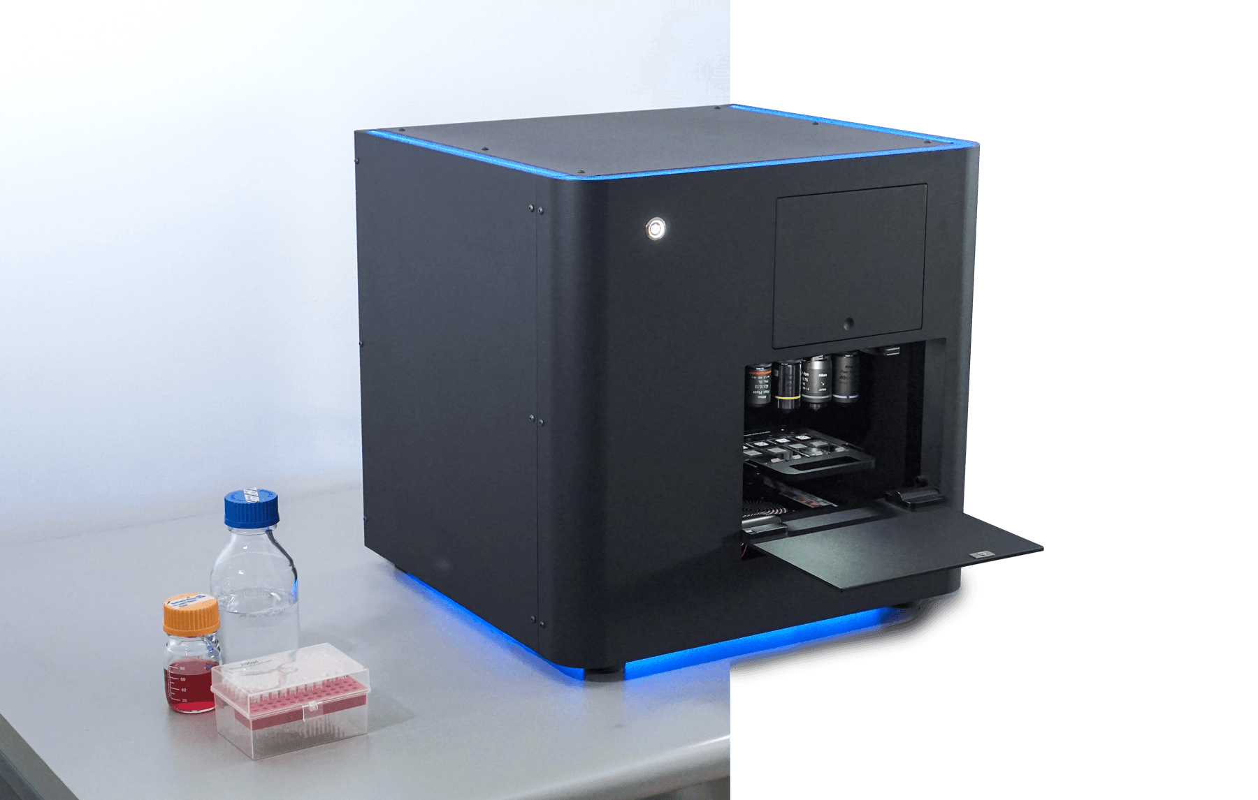
CytoCatch™ Imaging System
The CytoCatch™ Imaging System is an imaging-based device that was developed to scan, digitize, and view digitized images of slide specimens, in order to support clinical and research workflows. Thanks to its innovative illumination system, customization capacity, and intuitive user interface, the CytoCatch™ Imaging System digital slide scanner is capable of performing robust and high-speed scanning of fluorescent and non-fluorescent samples, enabling it to fulfill the most demanding needs of your clinical or research facility.
Advanced epi-illumination fluorescence microscopy
The CytoCatch™ Imaging System whole slide scanner light engine enables advanced epi-illumination fluorescence microscopy. Its 8-channel LED illumination delivers consistent, high-intensity excitation across the UV to far-red spectrum, with intelligent shuttering to minimize photo-bleaching.
Scan with multiple transillumination techniques
Scan your slide specimens with multiple transillumination techniques. From conventional brightfield to phase contrast and DIC, the imaging system adapts to virtually any cytology, histology, or hematology workflow without manual reconfiguration.
Features
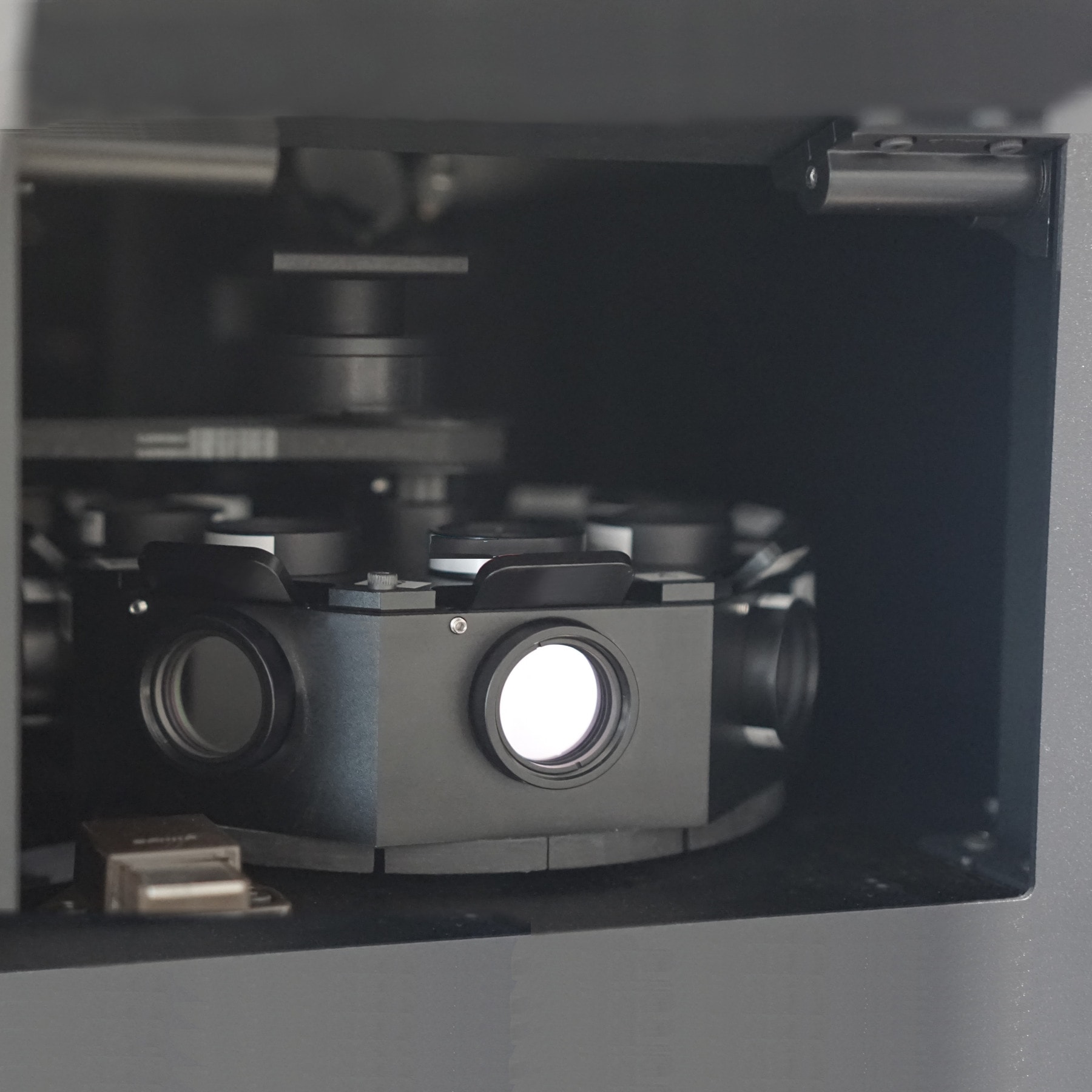
Maximum flexibility
Besides having an innovative LED 8 channel illumination system, the CytoCatch™ Imaging System digital slide scanner is prepared to accommodate up to 8 fluorescence filter cubes, which you can choose from our large selection of filter sets. This flexibility enables you to take advantage of the full range of the UV – far-red region, allowing the use of any commercially available stain or fluorophore in your staining process, and maximizing the signal to noise ratio of your samples.
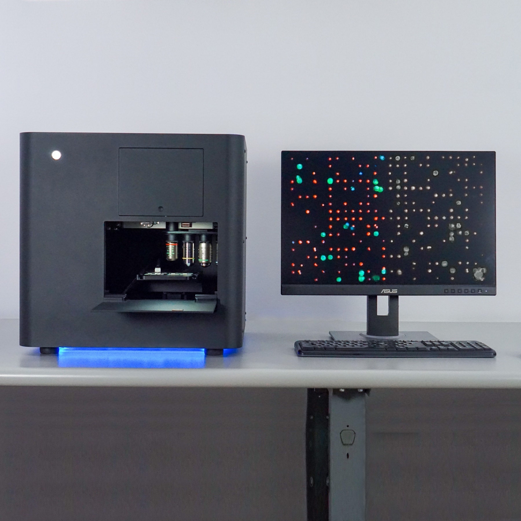
Dual camera
The CytoCatch™ Imaging System digital slide scanner is prepared to accommodate both a monochrome and color camera. The device automatically switches between these two depending on the employed microscopy technique, ensuring excellent image quality regardless of the nature of the sample.
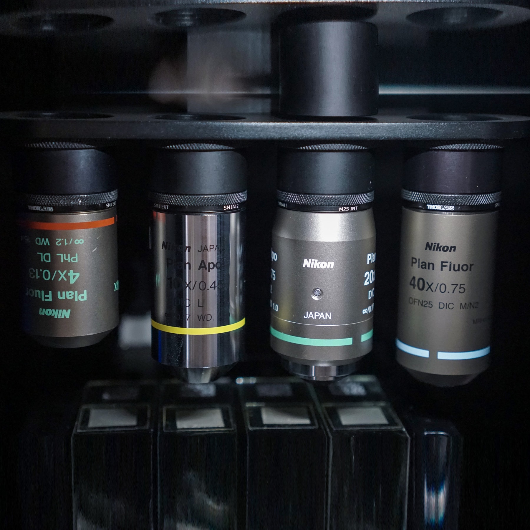
Multiple magnifications
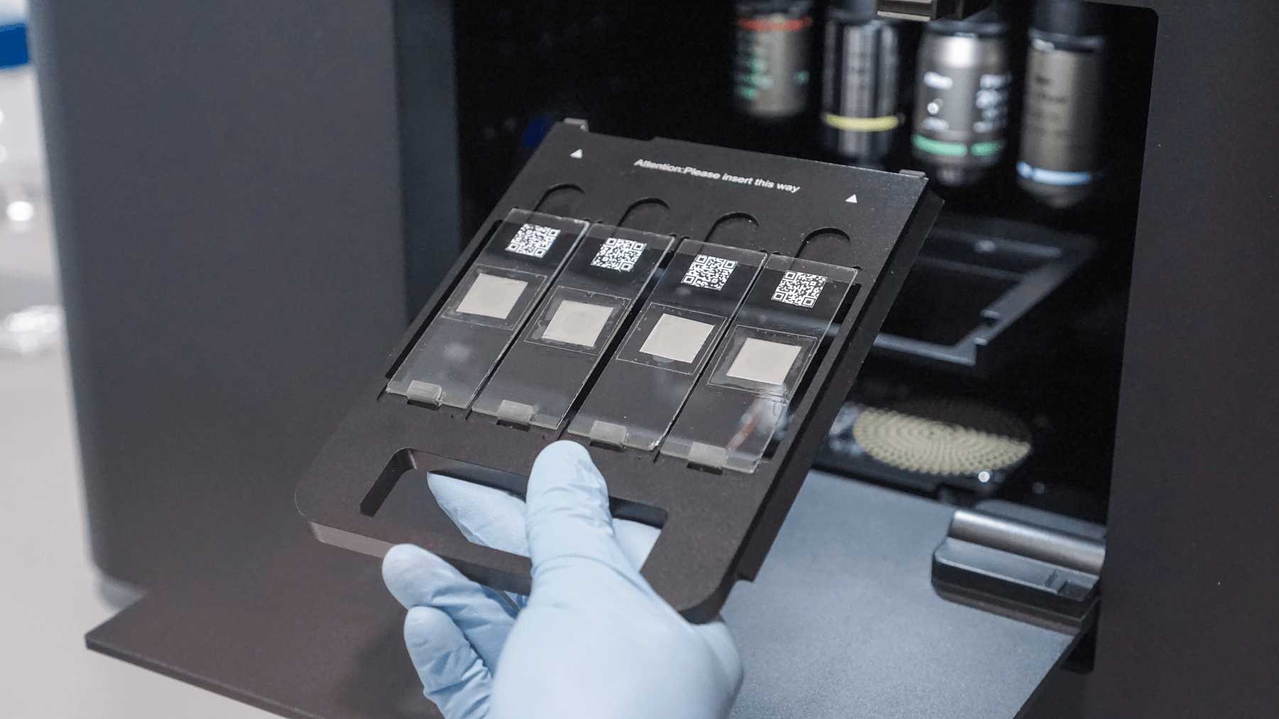
The CytoCatch™ Imaging System digital slide scanner is ideal for scanning whole slides at any magnification from 4× to 40×. The device is compatible with Nikon CFI objectives and has a 4-objective motorized nosepiece for automatic changing, enabling you to readily scan slides at their optimal magnification.
Unattended scans
The level of automation of the CytoCatch™ Imaging System digital slide scanner enables unattended scanning of samples. Slides are loaded on the device’s tray and scanned, eliminating slide-handling errors and improving reliability.
Always reliable
The CytoCatch™ Imaging System digital slide scanner has multiple sensors, auto-calibration routines, and built-in diagnostics to ensure that the device is always working properly, delivering high-quality scans.
Applications
Scan, digitize, and view digitized images of samples
Compatible with samples prepared with a variety of techniques, such as:
 Hematoxylin & Eosin staining (H&E)
 Immunohistochemistry (IHC)
 Immunocytochemistry (ICC)
 Immunofluorescence (IF)
 Special stains
 Cytology
 Frozen sections
Specifications
Technical overview
A complete technical snapshot of the CytoCatch™ Imaging System — optics, cameras, sample handling, and operating requirements.
Optics & illumination
- Light engine
- 8-channel LED, UV → far-red region
- Filter cubes
- Up to 8 fluorescence filter cubes
- Magnification range
- 4× to 40× (Nikon CFI objectives)
- Nosepiece
- 4-objective motorized, auto-switching
Cameras & imaging
- Cameras
- Monochrome + color, auto-switching
- Modes
- Brightfield, fluorescence, phase contrast
- Image output
- Whole-slide digital images, multi-channel
- Auto-calibration
- Built-in routines on every run
Sample handling
- Loading
- Multi-slide tray, unattended
- Slide format
- Standard 1×3 in (25×75 mm)
- Workflow
- Load → walk away → digitized slides
Operating environment
- Lighting
- Standard lab bench — no darkroom required
- Diagnostics
- Multi-sensor monitoring + self-test
- Software
- Integrated viewer + scan-protocol builder